Welcome to the Amira-Avizo Software Use Case Gallery
Below you will find a collection of use cases of our 3D data visualization and analysis software. These use cases include scientific publications, articles, papers, posters, presentations or even videos that show how Amira-Avizo Software is used to address various scientific and industrial research topics.
Use the Domain selector to filter by main application area, and use the Search box to enter keywords related to specific topics you are interested in.
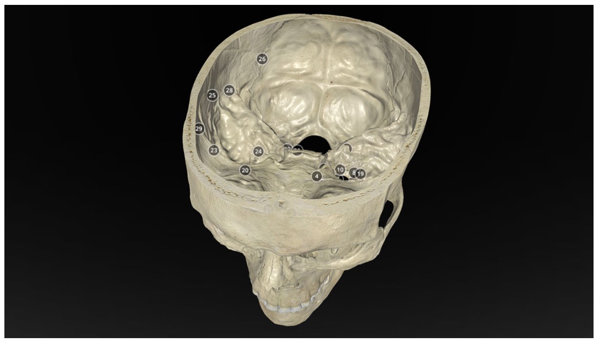
Operative Anatomy of the Human Skull: A Virtual Reality Expedition
Benjamin K Hendricks, MD Akash J Patel, MD Jerome Hartman Mark F Seifert, PhD Aaron Cohen-Gadol, MD, MSc, MBA
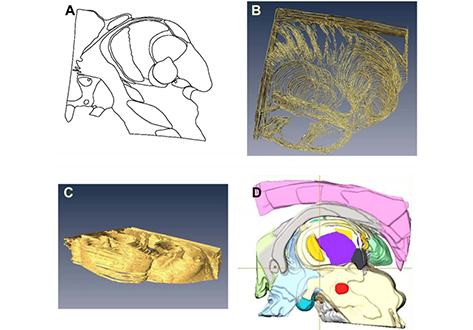
Classification and delineation of the motor-related nuclei in the human thalamus have been the focus of numerous discussions for a long time. Difficulties in finding consensus have for the most part been caused by paucity of direct experimental data on connections of individual nuclear entities. Kultas-Ilinsky et al. (2011, J Comp Neurol, 519:2811-2837) showed that distribution of the isoform 65 of glutamic acid decarboxylase (GAD65), the enzyme that synthesizes inhibitory neurotransmitt... Read more
Igor Ilinsky, Andreas Horn, Perrine Paul-Gilloteaux, Pierre Gressens, Catherine Verney, Kristy Kultas-Ilinsky
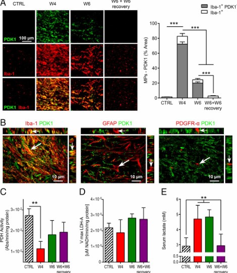
Proinflammatory mononuclear phagocytes (MPs) play a crucial role in the progression of multiple sclerosis (MS) and other neurodegenerative diseases. Despite advances in neuroimaging, there are currently limited available methods enabling noninvasive detection of MPs in vivo. Interestingly, upon activation and subsequent differentiation toward a proinflammatory phenotype MPs undergo metabolic reprogramming that results in increased glycolysis and production of lactate. Hyperpolarized (HP)
Caroline Guglielmetti, Chloé Najac, Alessandro Didonna, Annemie Van der Linden, Sabrina M. Ronen, and Myriam M. Chaumeil
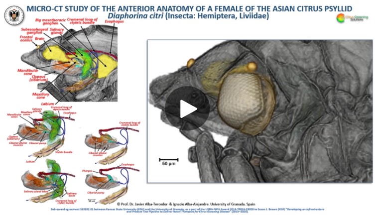
The Asian citrus psyllid (ACP), Diaphorina citri, is a harmful pest of citrus trees that transmits Candidatus Liberibacter spp. which causes Huanglongbing (HLB) (citrus greening disease); this is considered to be the most serious bacterial disease of citrus plants.
Here we detail an anatomical study of the external and internal anatomy (excluding the reproductive system) using micro-computed tomography (micro-CT). This is the first complete 3D micro-CT reconstruction o... Read more
Javier Alba-Tercedor, Wayne B. Hunter & Ignacio Alba-Alejandre
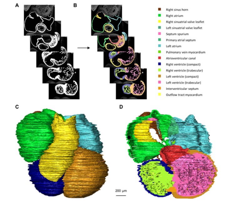
Quantified growth of the human embryonic heart
The size and growth patterns of the components of the human embryonic heart have remained largely undefined.
To provide these data, three-dimensional heart models were generated from immunohistochemically stained sections of ten human embryonic hearts ranging from Carnegie stage 10 to 23. Fifty-eight key structures were annotated and volumetrically assessed. Sizes of the septal foramina and atrioventricular canal opening were also measured. The heart grows exponentially throughout embr... Read more
Jaeike W. Faber, Jaco Hagoort, Antoon F. M. Moorman, Vincent M. Christoffels, Bjarke Jensen
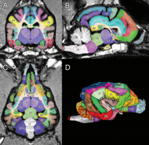
A detailed canine brain label map for neuroimaging analysis
Dogs have recently become an important model species for comparative social and cognitive neuroscience. Brain template-related label maps are essential for functional magnetic resonance imaging (fMRI) data analysis, to localize neural responses. In this study, we present a detailed, individual-based, T1-weighted MRI-based brain label map used in dog neuroimaging analysis. Methods: A typical, medium-headed dog (a 7.5-year-old
male Golden Retriever) was selected from a cohort of ... Read more
Czeibert Kálmán, Andics Attila, Petneházy Örs, Kubinyi Enikő, Kálmán Czeibert
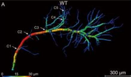
Accumulating evidence indicates the critical importance of cerebrovascular dysfunction in the pathogenesis of Alzheimer’s disease (AD). However, systematic comparative studies on the precise brain vasculature of wild-type and AD model mice are still rare. Using an image optimization method for analyzing Micro-Optical Sectioning Tomography (MOST) data, we generated cross-scale whole-brain 3D atlases that cover the entire vascular system from large vessels down to smallest capillaries at ... Read more
Xiaochuan Zhang, Xianzhen Yin, Jingjing Zhang, Anan Li, Hui Gong, Qingming Luo, Haiyan Zhang, Zhaobing Gao, Hualiang Jiang
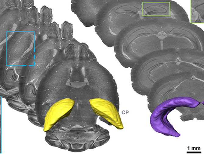
Precise Cerebral Vascular Atlas in Stereotaxic Coordinates of Whole Mouse Brain
Understanding amazingly complex brain functions and pathologies requires a complete cerebral vascular atlas in stereotaxic coordinates. Making a precise atlas for cerebral arteries and veins has been a century-old objective in neuroscience and neuropathology. Using micro-optical sectioning tomography (MOST) with a modified Nissl staining method, we acquired five mouse brain data sets containing arteries, veins, and microvessels. Based on the brain-wide vascular spatial structures and brain re... Read more
Benyi Xiong, Anan Li, Yang Lou, Shangbin Chen, Ben Long, Jie Peng, Zhongqin Yang, Tonghui Xu, Xiaoquan Yang, Xiangning Li, Tao Jiang, Qingming Luo and Hui Gong
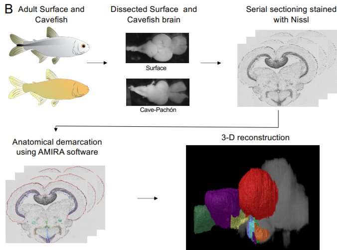
A shift in environmental conditions impacts the evolution of complex developmental and behavioral traits. The Mexican cavefish, Astyanax mexicanus, is a powerful model for examining the evolution of development, physiology, and behavior because multiple cavefish populations can be compared to an extant and ancestral-like surface population of the same species. Many behaviors have diverged in cave populations of A. mexicanus, and previous studies have shown that cavefish ha... Read more
Cody Loomis, View ORCID ProfileRobert Peuß, James Jaggard, Yongfu Wang, Sean McKinney, Stephen Raftopoulos, Austin Raftopoulos, Daniel Whu, Matthew Green, Suzanne E. McGaugh, Nicolas Rohner, Alex C. Keene, Erik R. Duboue
The Academic Medical Center (AMC) uses Amira to build a 3D Atlas for Human Embryology
The 3D Atlas of Human Embryology project was funded by the Academic Medical Center (AMC) in Amsterdam, the Netherlands, in 2009. Since then, over 75 students, under the supervision of embryologists of the Department of Anatomy, Embryology & Physiology, have contributed to this labor-int... Read more
Academic Medical Center